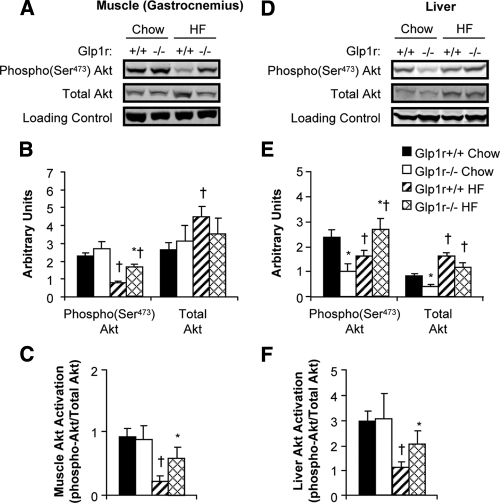
Figure 5.
Immunoblots for insulin signaling proteins after insulin clamp experiments. Representative immunoblots from gastrocnemius muscle (A) and liver (D) extracts are shown. Quantification of protein content for phosphorylated and total Akt (B and E) and Akt activation (C and F) is shown for chow-fed Glp1r+/+ (black bars), chow-fed Glp1r−/− (white bars), HF-fed Glp1r+/+ (striped bars) and HF-fed Glp1r−/− (diamond pattern bars) mice. GAPDH was used as a loading control. Data are shown as mean ± sem for 8–10 mice/genotype and diet. *P < 0.05 vs. Glp1r+/+, same diet; †P < 0.05 vs. Chow, same genotype.

