Figure 1.
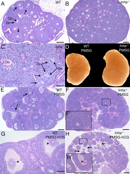
Histologic analysis of 3-wk-old WT and Inha−/− ovaries. Ovary from a 3-wk untreated WT mouse shows early antral follicle development (arrows) (A), whereas a 3-wk untreated Inha−/− ovary is larger and does not develop follicular antra to the same extent (B). C, Higher magnification of Inha−/− ovary, demonstrating uncoupled oocyte and granulosa cell growth (arrow) similar to studies of 12-d Inha−/− ovaries (11). D, Gross anatomy of 3-wk-old WT and Inha−/−ovaries injected for 44–46 h PMSG, demonstrating a larger size of the Inha−/− ovary. Three-week WT ovary 44–46 h after PMSG injection demonstrates preovulatory follicle development (E), whereas Inha−/− ovaries do not develop follicular antra (F). F′, Higher magnification of boxed area shown in F. G, Three-week-old WT ovary after 44–46 h PMSG and 6 h hCG containing cumulus oocyte complexes undergoing cumulus expansion (arrowheads). H, No cumulus expansion is visible in PMSG/hCG-treated Inha−/− ovaries (arrowhead). Boxed region in H is shown as a higher-magnification inset (H′). An unusual stromal component (arrows) between follicles is visible in PMSG/hCG-treated Inha−/− ovaries that is not seen in WT ovaries. Scale bars (A, B, E, F, and H), 200 μm. Scale bars (C), 50 μm. Scale bar (G), 100 μm. Oo, Oocyte; Gr, granulosa cells.

