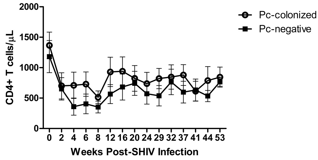
Figure 1.
Peripheral blood CD4+ T cell levels are not different for SHIV/Pc+ animals versus SHIV/Pc− animals. Peripheral blood mononuclear cells isolated from whole blood were stained with anti-CD4 antibody and analyzed by flow cytometry. Open circles represent SHIV/Pc+ animals and closed squares represent SHIV/Pc− animals. p = 0.79 by two-way repeated measures ANOVA for SHIV/Pc+ (n = 8) versus SHIV/Pc− group (n = 4).

