Figure 3.

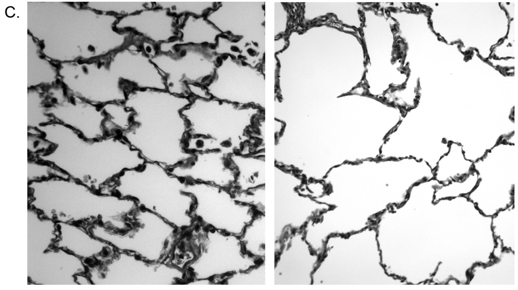
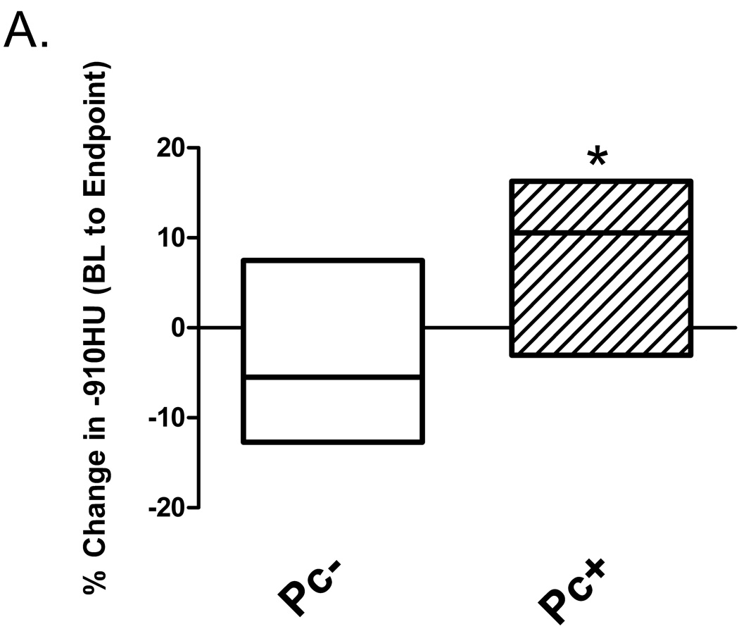
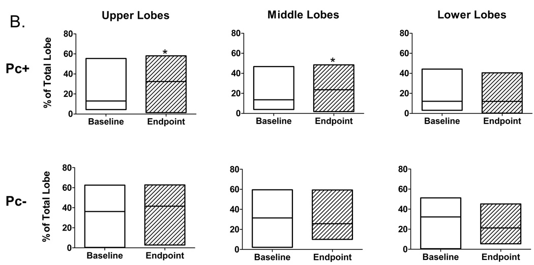
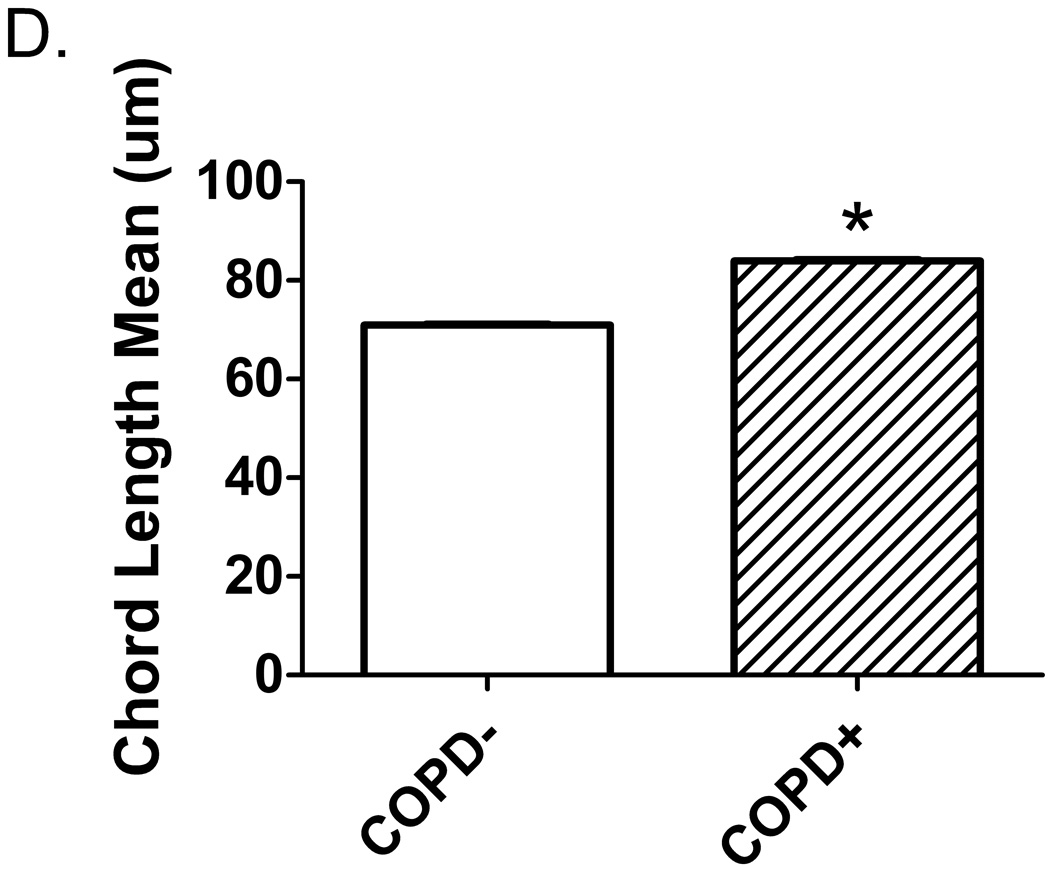
Pneumocystis colonization leads to an increase in the proportion of emphysematous tissue in the lungs. Quantitative computed tomography (CT) scans were performed at 20 cm H2O lung inflation pressure at baseline (BL) and post- SHIV infection. The cutoff mask of ≤ −910 Hounsfield units (HU) was used to assess amount of emphysematous lung tissue present at each scan. Boxes represent the range of values for the specified group with the median value represented by the line within the box. (a) Change in the proportion of emphysematous lung tissue for the animal groups; * p = 0.04 for SHIV/Pc− (n = 4) versus SHIV/Pc+ (n = 6†) animals by unpaired t test. (b) Comparison of the proportion of emphysematous lung tissue present at baseline and endpoint scan by lobe. For SHIV/Pc+ animals†: * p = 0.04 by paired t test, for the proportion of lung tissue that is emphysematous in both the upper and middle lobes for baseline versus endpoint scans; p = 0.78 by paired t test for proportion of lung tissue that is emphysematous in the lower lobe for baseline versus endpoint scans (n = 6). For SHIV/Pc− animals: p = 0.55, 0.80 and 0.11 by paired t test for proportion of lung tissue that is emphysematous in the upper, middle and lower lobes respectively for baseline versus endpoint scans (n = 4). (c) Representative hematoxylin-stained lung tissue sections from SHIV-infected monkeys; left: SHIV/Pc− and right: SHIV/Pc+. (d) Chord length analysis (mean ± SEM) of airspaces for animals exhibiting clinical type (≥ 12% decline in pulmonary function from baseline level) obstruction (COPD+, n = 5) versus non-obstructed animals (COPD−, n = 7), *p = 0.0001.
†Two SHIV/Pc+ animals were not included in either the pre- or post-infection analyses because baseline scans were not performed. Both of these animals developed airway obstruction based on pulmonary function testing.


