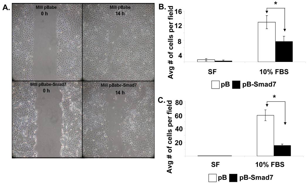
Figure 3. Smad7 overexpression suppresses migration and invasion of MIII cells.
A. Representative light microscope images of wound healing assays for MIIIpB and MIIIpBSmad7 cells to evaluate their migration rate into the cell-free area. B. Chemotaxis assay. Cells that migrated through the 8µm pore-containing membrane of the transwells were stained with propidium iodide (PI) and counted. C. Matrigel invasion assay. Cells that invaded through matrigel were stained with Trypan blue and counted. All results are presented as the average of cells counted in ten fields per condition.

