
FIG. 2.
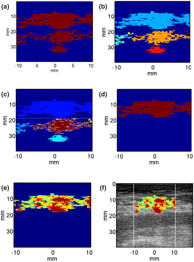
Method for automatic identification of pixels corresponding to extravasated blood. (1) A binary mask is developed by thresholding on mean variance of displacement measured from 20 to 60 ms of the acquired ARFI displacement tracking ensemble (panel (a)). (2) Selected pixels are spatially grouped into clusters using ‘bwlabel’ in MATLAB with 8 connected objects (panel(b)). (3) The cluster with the highest mean variance of the 2nd derivative of correlation coefficient (CC) is identified (outlined); it and all other clusters at or below its axial position are rejected as luminal blood (panel (c)). (4) Pixels with peak displacements >4 μm in the remaining clusters are identified as extravasated blood pixels (panel (d)). ARFI PD values are mapped to the extravasated blood pixels (panel (e)) and hybrid ARFI/B-Mode images are rendered (panel (f)).