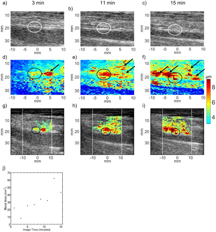
FIG. 3.

Serial B-Mode, ARFI peak displacement (PD), and hybrid ARFI/B-Mode images of the femoral arteriotomy in a 76 year-old female volunteer at 3 (left column), 11 (middle column) and 15 (right column) min following sheath removal. Hemostasis was achieved by manual compression alone. In B-Mode images (top row, panels (a), (b) and (c)), the arteriotomies (circled) are not readily apparent, and there is no indication of bleeding. In raw ARFI PD images (middle row, panels (d), (e) and (f)), the arteriotomy is notable as a localized region of relative high tissue displacement in the near arterial wall (circled). Relatively large ARFI PDs in the soft tissue above the artery with high displacement variance are suggestive of extravasated blood (arrows). Hybrid ARFI/B-Mode images (third row, panels (g), (h) and (i) map ARFI PDs in positions of high displacement variance into B-Mode images and more clearly suggest a growing pool of extravasated blood. The hybrid images support bleeding detection but some arterial wall detail at 3 min follow sheath removal (panel (g), circled) is sacrificed. A scatter plot of estimated extravasated blood area versus time (panel (j)) shows consistently increasing area and suggests that hemostasis is not achieved within the 15 min serial imaging period.
