
FIG. 4.
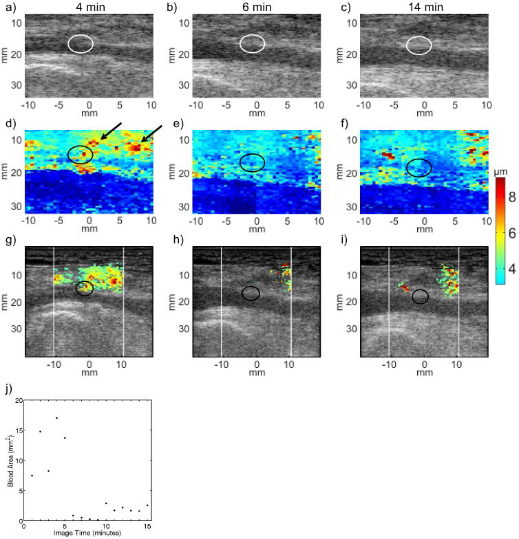
Serial B-Mode, ARFI peak displacement (PD), and hybrid ARFI/B-Mode images of the femoral arteriotomy in a 60 year-old female volunteer at 4 (left column), 6 (middle column) and 14 (right column) min following sheath removal. Hemostasis was achieved by manual compression augmented the pGlcNAc fiber based dressing. B-Mode images (top row, panels (a), (b) and (c)) indicate neither the arteriotomies (circled), nor whether hemostasis has been achieved. Raw ARFI PD images (middle row, panels (d), (e) and (f)) show a focal region of relatively large peak displacement at the arteriotomy (circled) at 4 min following sheath removal (panel d). Relatively large ARFI-induced displacement suggests extravasated blood at the 4 min time point but not at the 6 and 14 min time points (panels (e) and (f), respectively). Hybrid ARFI/B-Mode images (third row, panels (g), (h) and (i)) more clearly show that the estimated area of extravasated blood decreases at 6 min. A scatter plot of estimated extravasated blood area versus time (panel (j)) shows a drop in area at 6 min that is sustained throughout the duration of imaging, suggesting hemostasis onset at 6 min.