Figure 4.

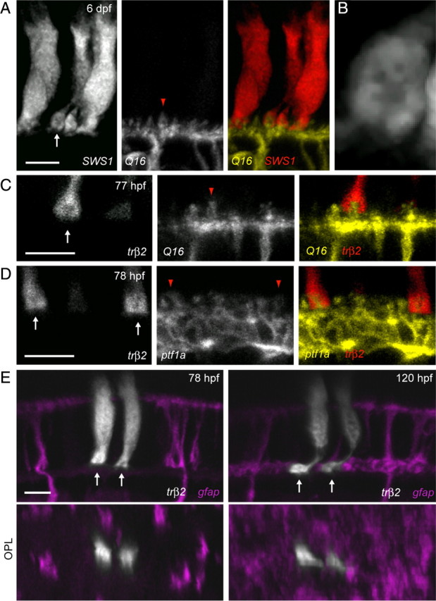
Cone pedicle invagination precedes MG process arrival in the OPL. A, Cone photoreceptors expressing tdTomato driven by the SWS1 promoter in the background of the Q16 line labeling ON bipolar cells. Arrow points to a cone pedicle. B, High magnification of the cone terminal in A (arrow) reveals the presence of a dark region within the pedicle, suggestive of the location of the invaginations of postsynaptic HC and bipolar cell dendrites. C, Insertion of a bipolar cell (yellow, Q16) dendritic tip (red arrowhead) into a cone photoreceptor (red, trβ2) pedicle (arrow) at an age (77 hpf) where MG processes are absent from the OPL. D, Insertion of HC (yellow, ptf1a) dendritic tips (red arrowheads) into cone photoreceptor (red, trβ2) pedicles (arrows) at an age (78 hpf) where MG processes have not infiltrated the OPL. E, A pair of cone photoreceptors (grayscale) transiently expressing tdTomato at 78 hpf demonstrate invaginations (dark spots) in their pedicles (arrows) before the arrival of MG processes (magenta) in the OPL. Orthogonal rotations (OPL) clearly show that cone pedicles were not contacted by MG processes at this stage. The same cones and MGs imaged at an age when MG processes surround the cone pedicles (right). Cones were labeled by injecting trβ2:tdTomato plasmid into the transgenic line gfap:GFP. Scale bars, 5 μm.
