Figure 7.
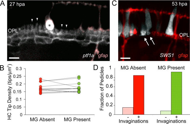
MG processes are not required for HC dendritic tip or cone photoreceptor pedicle stability. A, Six MGs were ablated at 4 dpf in a triple transgenic line labeling HCs (grayscale, ptf1a:G4VP16; UAS:MYFP) and MGs (pink, gfap:GFP). Examples of HC dendritic tips remaining in areas that lack MG processes are indicated by arrowheads. Asterisk indicates swollen debris of apical MG process. B, HC dendritic tip densities were compared within the field of view for regions where MGs were absent (red dots) and regions where they were present (green dots). Lines connect measurements from the same field (an ablation experiment). Analysis was performed only in regions large enough to contain at least five dendritic tips, and were pooled across all time points after ablation (1–4 d after ablation). n = 13 ablated regions. C, Six MGs were ablated at 4 dpf in retinas where UV cones transiently expressed tdTomato (grayscale) in the gfap:GFP transgenic line (red). UV-cone photoreceptor invaginations (dark spot within the pedicles) are indicated with arrows. D, Fraction of UV-cone photoreceptor pedicles that collapsed (−) or remained invaginated (+) in areas lacking MG processes (red, n = 26) or where MG processes were present (green, n = 38). Scale bars, 5 μm.

