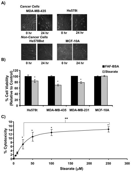
Figure 2. Stearate decreased the viability of the human breast cancer cells.
A) Stearate induced morphological changes of the breast cancer cells but not the non-cancerous cells 24 hours post treatment. B) Twelve hours post-treatment, stearate decreased the viability of the breast cancer cells but not the non-cancerous MCF-10A cells using the trypan blue exclusion assay (n=2 performed in triplicate; *p<0.005 by the Student's t-test). C) To confirm the decrease in cell viability, cytotoxicity induced by stearate 12 hours post-treatment was estimated by measuring lactate de-hydrogenase activity in the cell culture media. Stearate decreased the viability of the cancer cells in a dose dependent manner. (n=3 performed in triplicate; *p<0.04 compared to FAF-BSA, **p<0.02 by ANOVA).

