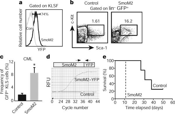
Figure 2. The presence of constitutively active Smo increases the frequency of CML stem cells and accelerates disease.
a, Analysis of YFP fluorescence, which reflects SmoM2 expression in control (con) and SmoM2 KLSF cells (n = 4). b, Analysis of CML stem cells (GFP+ KLS) in mice transplanted with BCR–ABL1-infected control (left) and constitutively activated Smo (SmoM2, right) KLSF cells. c, The average percentage of CML stem cells in mice receiving BCR–ABL1-transduced control (n = 4) and SmoM2 KLSF cells (n = 12). Error bars are s.e.m., *P = 0.048. d, rtPCR analysis of SmoM2 expression in CML stem cells. e, Survival curve of mice receiving BCR–ABL1 transduced control (solid line) or SmoM2 KLSF cells (dashed line; n = 2, 16 mice, P = 0.0082).

