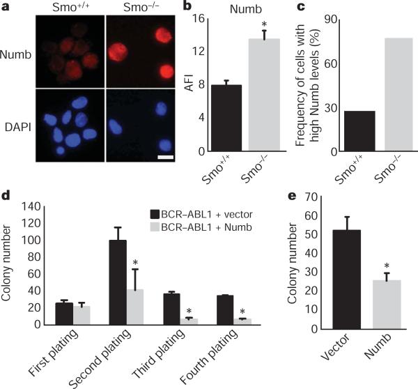
Figure 3. Loss of Smo increases frequency of cells with high levels of Numb and contributes to decreased CML growth.
a, CML stem cells from Smo+/+ and Smo−/− leukaemias were stained with Numb (red, top panel) and 4,6-diamidino-2-phenylindole (DAPI; blue, bottom panel). Scale bar, 10 μM. b, The average fluorescence intensity (AFI) was determined by dividing the overall mean fluorescence intensity by the area of the cell (P = 0.002). c, The frequency of cells with high expression levels of Numb was calculated by designating cells above a mean fluorescence intensity value of 1,000 as high expressors (n = 3, using either CML KLS or CML c-Kit+ cells). d, KLSF cells were infected with MSCV-BCR-ABL1-IRES-GFP and either vector MSCV-IRES-YFP or MSCV-Numb-IRES-YFP. GFP and YFP double-positive cells were plated in methylcellulose, colonies were counted and cells were serially replated (n = 2, *P < 0.006). e, CML KLS cells were infected with viruses expressing control vector or Numb IRES-YFP. Infected cells were sorted and plated in methylcellulose (n = 2, P = 0.03). Error bars in b–e are s.e.m.

