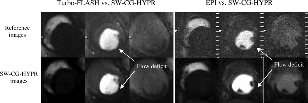
Figure 4.
Comparison of conventional method (first line) and SW-CG-HYPR (bottom line). Similar signal changes in the left ventricle and myocardium were observed between the SW-CG-HYPR and the conventional methods. The spatial resolution and the apparent SNR of the SW-CG-HYPR images were higher than the conventional images. The myocardial territories supplied by the LCX artery is better delineated in SW-CG-HYPR images than in the reference images.

