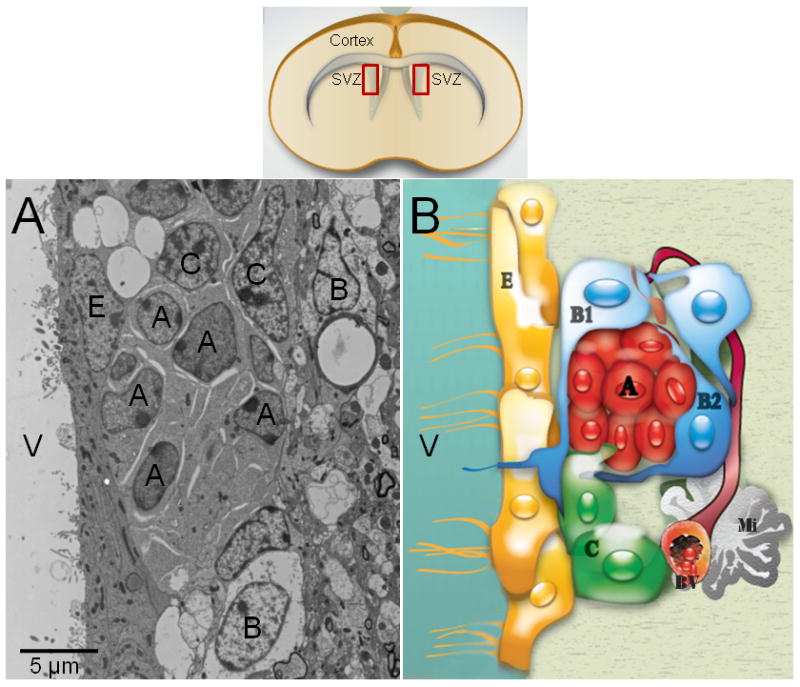
Fig. 1. The adult subventricular zone in rodent.

This neurogenic niche has been well-characterized by electron microscopy (A). Type-A cells (migrating neuroblasts) have an elongated cell body with one or two processes, profuse lax chromatin with small nucleoli (2 to 4), a scarce dark cytoplasm, abundant free ribosomes, microtubules oriented along the long axis of the cells, and nuclei occasionally invaginated. Their cytoplasmic membrane showed cell junctions intercalated with large intercellular spaces. Type-B cells have a light cytoplasm with a few ribosomes, extensive intermediate filaments and nuclei are typically invaginated. Their cell profiles are irregular that filled intercellular spaces between neighboring cells. Type-C cells are large and semi-spherical; their nuclei contain deep invaginations, lax chromatin occasionally clumped and large reticulated nucleoli. Type-C-cell cytoplasm is more electron-lucent than Type-A cells, but more electron-dense than Type-B cells, because it contains a few ribosomes and no intermediate filaments. Schematic drawing of the adult SVZ shows the cell organization of this region (B). Ependymal cells (Type-E cells) formed an epithelial monolayer that separates the SVZ from the lateral ventricles. These cells have spherical nuclei with lax chromatin, lateral cytoplasmic processes heavily interdigitated with apical junction complexes. The cell membrane contacting the ventricle contains microvilli and cilia, and their cytoplasm is electron-lucent with many mitochondria and basal bodies in the apical pole. A: Type-A cell; B: Type-B cell; C: Type-C cell cell; E: Ependymal cell; BV: Blood vessel; Mi: Microglia cell; V: Ventricle.
