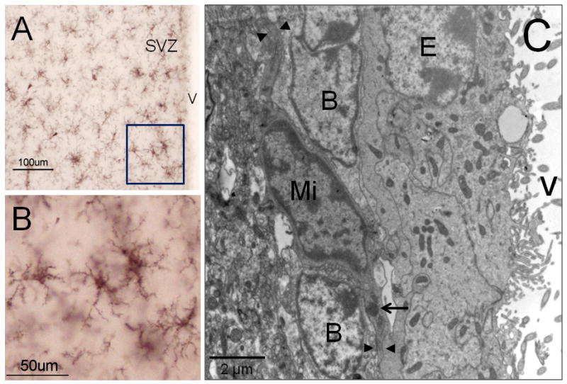
Figure 3. Microglia cells in the rodent SVZ.

Iba-1 immunocytochemistry to detect microglia cells by light microscopy (A-B). In their “resting” stage microglia displays multiple thin branches, which confer a ‘bushy’ morphology to these cells. By electron microscopy (C), microglia (Mi) shows a typical dark nucleus and electrodense corpus in their cytoplasm (arrow). Frequently, microglial cytoplasmic expansions (arrow heads) are in close contact with Type-B cells. B: Type-B cell; E: Ependymal cell; V: Ventricle.
