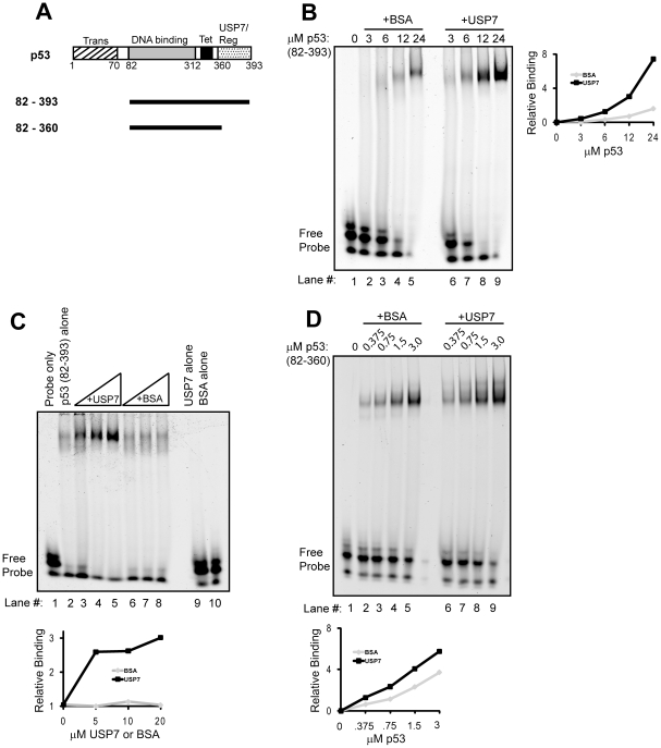
Figure 1. Effect of USP7 on the DNA binding activity of p53 in vitro.
(A) Schematic representation of the p53 proteins used in this study showing the transactivation (Trans), DNA binding core, tetramerization (Tet) and USP7-binding and regulatory (USP7/Reg) regions. (B) EMSA showing titration of latent p5382–393 in the presence of 20 µM of BSA negative control (lanes 2–5) or 20 µM USP7 (lanes 6–9). (C) EMSA performed with fixed amount (12 µM) of p5382–393 and with 5 µM, 10 µM or 20 µM of USP7 (lanes 3–5) or BSA (lanes 6–8). Incubation of 20 µM of USP7 alone or BSA alone with labeled probe in the absence of p53 is also shown (lanes 9 and 10). (D) EMSA showing titration of active p5382–360, in the absence (lanes 2–5) or presence of USP7 (lanes 6–9). Quantification of the discreet shifted bands for parts B,C and D are shown in the graphs, with USP7 in black and BSA in grey.

