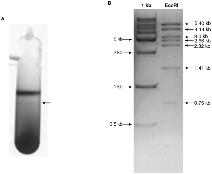
Figure 7. Confirmation of the presence of a plasmid in C. carboxidivorans strain P7T.
A) Isolation of plasmid p19 from strain P7T by standard cesium chloride gradient. Visualization of the two bands of DNA with UV illumination. Arrow indicate the lower band which contains the closed circular plasmid DNA. B) Digestion of the plasmid DNA by EcoRI. 1 kb ladder was used. Sizes are indicated on the left (ladder) and on the right (plasmid DNA).

