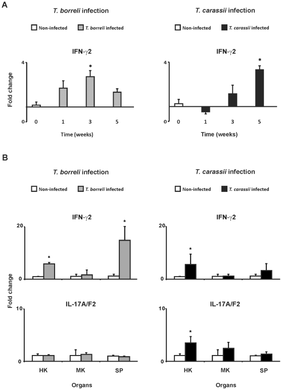
Figure 7. IFN-γ2 and IL-17A/F2 gene expression during T. borreli- and T. carassii-infection.
Gene expression was measured at different time points post-infection in peripheral blood leukocytes (PBL) (A) or at a fixed time point (3 weeks) in head kidney (HK), mid kidney (MK) and spleen (SP) (B). Carp were injected (i.p.) with a dose of 10000 parasites per fish, PBS-injected individuals served as negative controls. mRNA levels of IFN-γ2 and IL-17A/F2 were normalized against the house keeping gene 40S ribosomal protein S11 and expressed as fold change relative to non-infected fish. Data points represent averages + SD of n = 3 non-infected fish (indicated as week 0) and n = 3 infected fish per time point. Symbol (*) represents a significant (P≤0.05) difference compared to non-infected fish.

