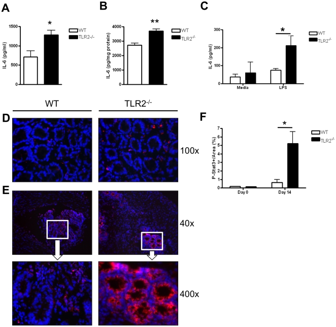
Figure 5. TLR2−/− mice have increased IL-6 and STAT3 activation during early intestinal tumorigenesis.
Panels A–C: IL-6 production in WT and TLR2−/− mice at day 14 of AOM-DSS treatment was measured by ELISA. (A) Serum levels of IL-6 (n = 8 WT, n = 9 TLR2−/−). (B) IL-6 concentration in colon homogenates was normalized to concentration of protein in the tissues (n = 3 WT, n = 5 TLR2−/−). (C) IL-6 secretion from isolated colonic lamina propria cells treated with LPS (1 µg/ml) for 6 h (n = 3–5). (D) Immunofluorescent stains of phospho-Stat3 in colonic tissues of WT and TLR2−/− mice at baseline. Original magnification 100x. (E) Immunofluorescent staining of phospho-Stat3 in colonic tissues of WT and TLR2−/− mice treated for 14 days with the AOM-DSS regimen. Top panel original magnification 40x, bottom panel original magnification 400x. (F) Quantification of phospho-Stat3 intensity measured in ACF from greater than five focus fields in at least four slides per animal (n = 5 WT and n = 5 TLR2−/−). All tests were performed using 95% confidence intervals. Data are expressed as means ± SEM. * = p<0.05, ** = p<0.01, *** = p<0.001.

