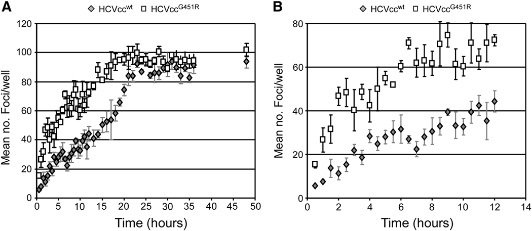
Fig. 4. Mutations that alter HCV particle density affect viral infection initiation kinetics.
Huh7 cells were plated in 96-well plates, infected with ~ 100 FFU of HCVccwt (grey diamonds) or HCVccG451R (open squares) and at indicated time points p.i., inoculum was removed from triplicate wells, cells were washed twice with 1X PBS and overlaid with 10% cDMEM containing 0.25% methylcellulose. Forty-eight hours p.i., HCV foci were visualized by HRP-based immunocytochemistry staining using an anti-NS5A antibody and are expressed as mean number (no.) of foci detected/well ± stdev for triplicate samples. Linear trend lines are depicted to help illustrate the underlying difference between the two viruses.

