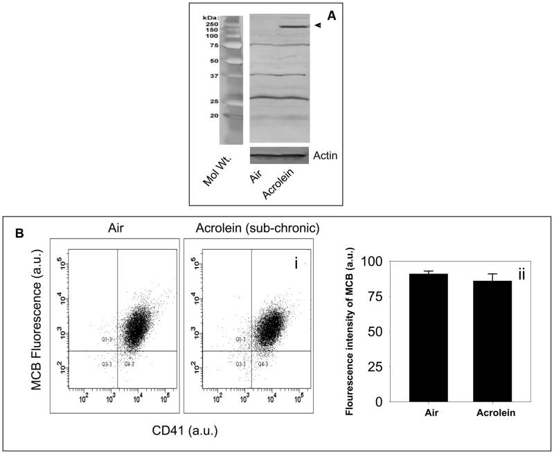
Figure 2. Increased protein-acrolein adduct formation and no change in GSH levels in acrolein-inhaled mice.
Panel A shows the representative Western blot of lysates of washed platelets obtained from mice exposed to acrolein (1 ppm/6h/day for four days; n=3) or filtered air (n=3), and probed with anti-acrolein-KLH antibody. Panel B shows the flow cytometric analysis of intracellular reduced glutathione levels in the platelets of mice exposed to acrolein (1 ppm/6h/day for four days; n=5) or filtered air (n=5). Washed platelets were labeled with monochlorobiamine (MCB) and mean fluorescence intensity of MCB was measured at excitation and emission wavelengths of 394 and 490 nm respectively. Representative illustration of fluorescence intensity of MCB in platelets isolated from control (air-exposed) and acrolein-exposed (1 ppm for 6h/day for four days) mice is shown in panel (i) and the group data is shown in panel (ii) as percent CD41-MCB double positive cells from total events. Values are mean ± SEM.

