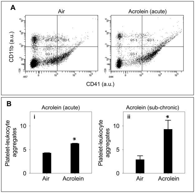
Figure 5. Acrolein exposure increases platelet-leukocyte adduct formation.
Panel A shows representative tracings of flow cytometric analysis of platelet-leukocyte aggregate formation in the whole blood of acrolein-exposed mice. Mice were exposed to acrolein (5 ppm for 6h) or filtered air and platelet-leukocyte aggregate formation was measured in the whole blood by FACS analysis, using PE-labeled anti-CD41 and PerCP-cy5.5-labeled anti CD11b antibodies. FSC versus SSC scales are shown to distinguish platelets from other cells and cell debris. Representative illustration in quadrent 2.1 shows platelet-leukocyte aggregates of double positive cells in control (air) and acrolein-exposed mice. Group data (n=8/group) for the platelet-leukocyte aggregates in acrolein-inhaled mice are shown in panel Bi. Panel Bii shows the group data for platelet-leukocyte aggregate formation in mice following sub-chronic (1 ppm for 6h/day for 4 days; n=8) exposure to acrolein or filtered air (controls; n=8). Values are mean ± SEM. *P ≤ 0.01 versus controls.

