
Figure 2. Effect of DON on VM-M3 cells in vitro.
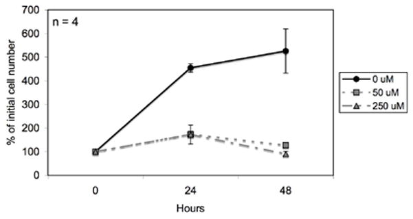
VM-M3/Fluc cells were seeded in DMEM in 24-well plates as described in the Materials and Methods and treated with DON (50 μM and 250 μM). Cells were imaged every 24 hrs using the Xenogen IVIS System. Data are expressed as the mean percent increase in cell number relative to the 0 hr time point ± 95% C.I. of 4 independent samples per group.