
Figure 5. Influence of DON or CR on liver histology.
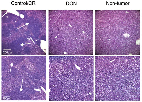
Removed livers were stained with haematoxylin and eosin (H & E) as described in Materials and Methods. Arrows indicate secondary tumor lesions in the control and CR group. Images are shown at 100X (top panel) and 200X (bottom panel).