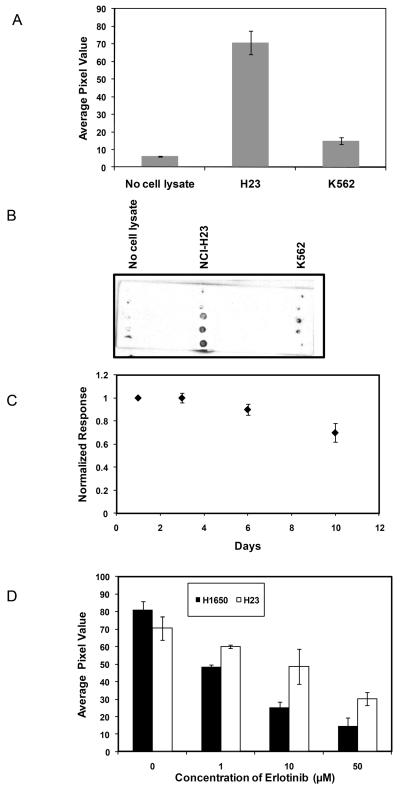
Figure 2.
Specificity of immobilized peptide phosphorylation by EGFR. (A) H23 lysate, K562 lysate, and no lysate control were assayed for EGFR activity. ECL values represent the extent of substrate phosphorylation. (B) Typical image of phosphorylation of immobilized peptide after incubation with H23, K562 cell lysates and no lysate control for 1 hr. (C) Comparison of substrate phosphorylation following incubation in PBS buffer for 1-10 days. (D) Comparison of the effects of erlotinib on the phosphorylation of immobilized peptide by H23 (cell with wild type EGFR) and H1650 (mutant EGFR) cell lysate. Each data point represents the mean of three independent experiments in which each experiment contained three replicates. The error bars represent s.e.m. (n=3).

