
Fig. 2.
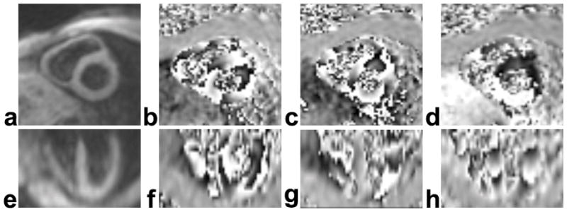
Example magnitude- (a,e) and phase-reconstructed 3D spiral cine DENSE images (b–d, f–h) of the heart at end-systole. The 3D volume was oriented along the principal axes of the LV. Online image reconstruction depicted short-axis planes of the LV, as shown in the magnitude- (a) and phase-reconstructed (b–d) images in the upper row. The lower row contains corresponding data reformatted offline in a long-axis four-chamber view. The images in (b) and (f) were encoded for displacement in the horizontal direction of the short-axis plane, in (c) and (g) were encoded for displacement in the vertical direction of the short-axis plane, and in (d) and (h) were encoded for displacement in the longitudinal direction.