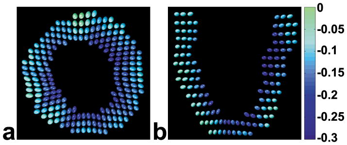
Fig. 5.
Ellipsoid visualization of 3D end-systolic LV function for one volunteer. In short-axis (a) and long-axis (b) views reconstructed from full volumetric 3D data sets, displaced (relative to their end-diastolic positions) ellipsoids represent both motion and 3D strain. For 3D strain, the lengths and orientations of the principal axes of the ellipsoids are determined by the lengths and orientations of the principal strains. Also, the ellipsoids are color coded according to Ecc. The direction of the first principal strain generally points toward the center of the LV. Transmural gradients of strain are evident, with greater radial lengthening and circumferential shortening occurring in the subendocardium vs the subepicardium. Multiphase data are displayed as corresponding ellipsoid movies in online supplemental data.

