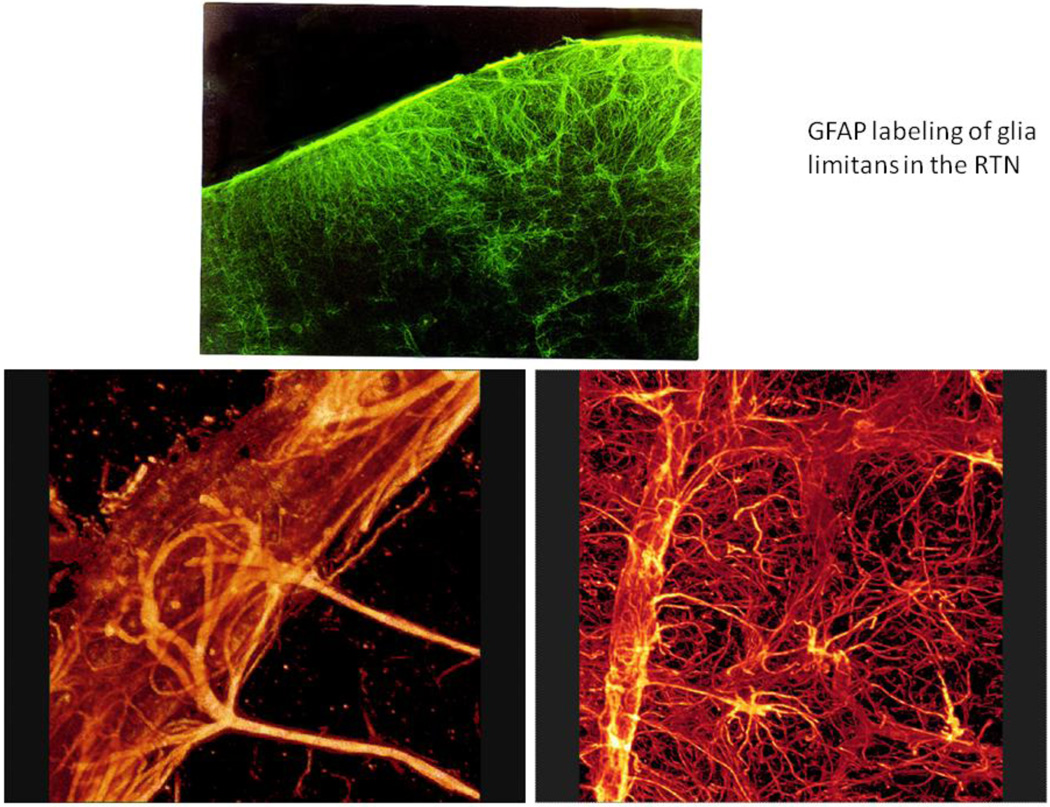
Figure 1.
In the upper panel, the glia limitans on the ventral surface of the medulla (up) has been stained immunohistochemically using an antibody directed against glial fibrillary acid protein (GFAP). Note the density of astrocytes near the ventral surface. The lower panels demonstrate the dense investment of blood vessels by astrocytic endfeet and the extensive filling of the neuropil in the ventral medulla by astrocytic processes. Astrocytes are more sparsely distributed in the dorsal medulla; for example, in the NTS there are relatively few astrocytes (Erlichman et al., 2004).

