Figure 1.

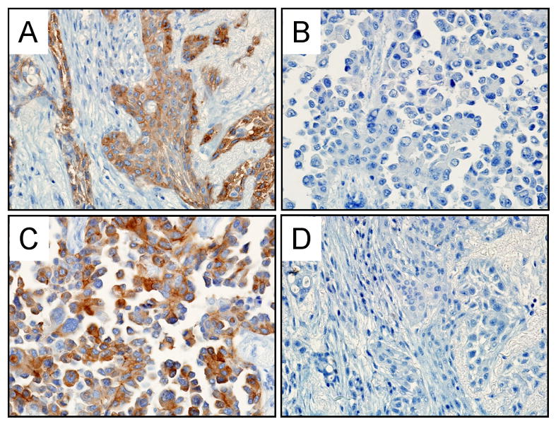
Representative pictures of EGFR expression by immunohistochemical staining (40× magnification). A. DNA sequence E746-A750 deletion specimen stained with the E746-A750 specific antibody. Immunoreactivity is positive in the tumor cells but absent in non-tumor cells. B. DNA sequence L858R point mutation case. The tumor cells show no immunoreactivity with the E746-A750 specific antibody. C. L858R point mutation tumor showing tumor cells positive for L858R specific antibody. D. E746-A750 mutation case displaying no immunoreactivity in the tumor or non-tumor cells with the L858R specific antibody.
