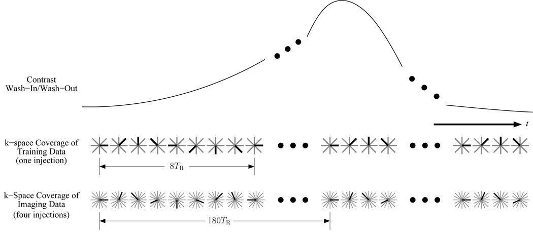
Figure 2.
The k-space sampling over time for the proposed method. The dark radial is the one acquired for that TR, while the light gray radials represent the full set. The training data cover 8 equally spaced angles and are incremented by 45° every TR. The imaging data cover 720 equally spaced angles that are divided into 4 unique sets for acquisition during separate contrast injections. The imaging data’s angle step size is 66.5° every TR.

