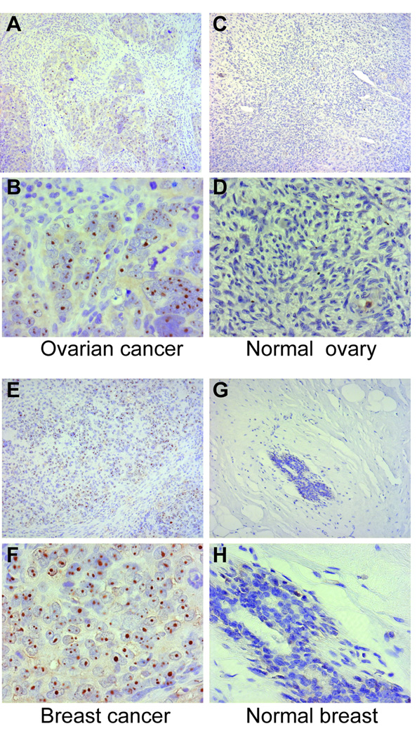
Figure 3. RNA polymerase III staining is prominent in cancerous ovary and breast tissue from scleroderma patients compared to normal ovary and breast. Paraffin sections from cancerous ovary (panels A and B) and breast (panels E and F) from scleroderma patients with cancer, as well as normal ovary (panels C and D) and normal breast (panels G and H) were stained with antibodies against RNA polymerase III as described in the methods section.
In all panels, the brown color represents RNA polymerase III staining, with nuclei in blue (Mayers’ hematoxylin counterstain). Magnifications are 10× (upper panels of each set - panels A, C, E and G) and 40× (lower panels of each set - panels B, D, F and H). In each set, the 40× panel is a magnification of part of the field shown at10×. The cancer sections shown were from subjects # 4 (ovarian cancer) and # 42 (breast cancer).

