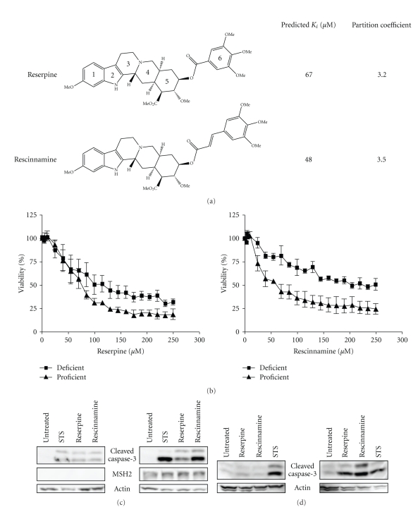
Figure 1.
(a) Structures of reserpine and rescinnamine with their binding constants and calculated partition coefficients (see Methods for details). (b) MTS cell survival assay shows MSH2-dependent cell viability after concentration-dependent reserpine (left) and rescinnamine (right) treatments. MSH2-deficient cell response is shown with squares, proficient response with triangles. (c) Western blot shows caspase-3 cleavage in Hec59 (MSH2-deficient; left panel) and Hec59(2) (MSH2-proficient; right panel) cells upon treatment with reserpine (85 μM) and rescinnamine (60 μM). The 17 and 19 kDa cleavage products of caspase-3 are depicted. Staurosporine (STS) was used as a positive control; actin was used as a loading control. (d) Western blot displays caspase-3 cleavage in DLD1 (MSH6-deficient; left panel) and DLD1(2) (MSH6-proficient; right panel) cells treated with reserpine and rescinnamine.

