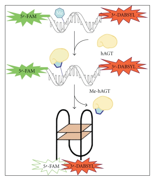
Figure 1.
Scheme of fluorescence-based hAGT assay. The substrate is the thrombin-binding aptamer modified by an O6-methylguanine (Me) in extended conformation, with a fluorophore and quencher in the opposite ends of the sequence. The refold of the G-quadruplex structure of TBA is dependent on the removal of the methyl adduct by alkyl-guanine-DNA-O6-alkyltransferase (hAGT). This repair moves the quencher and the fluorophore molecules in close proximity and blocks fluorescence.

