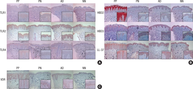
Fig. 1.
The immunohistochemical studies of psoriasis lesional skin (PP) and perilesional normal skin (PN), atopic dermatitis lesional skin (AD), healthy normal control skin (NN). (A) Expression of TLR1, TLR2 and TLR4. (B) Expression of HBD2, HBD3 and LL-37. (C) Expression of VDR (×100, inset: ×400).

