Figure 2.

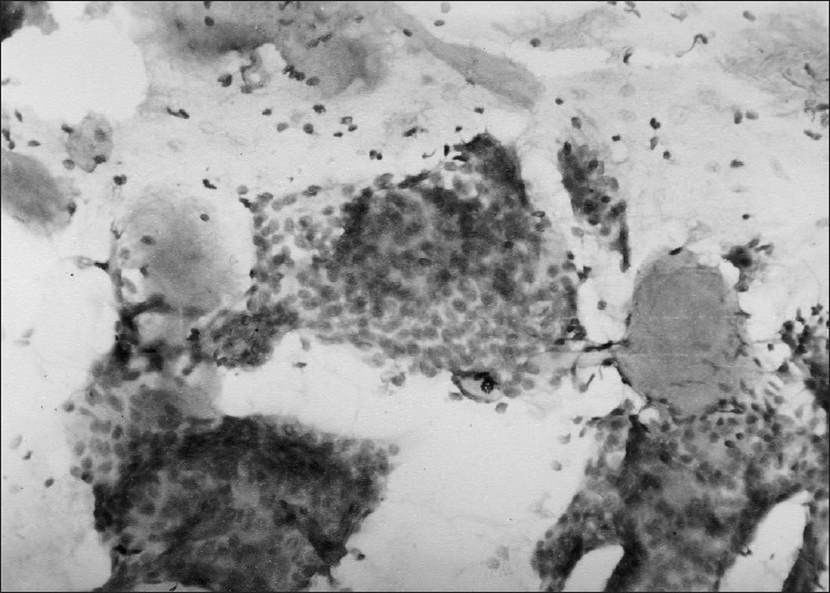
FNAC smear showing spherical globules of basement membrane material and hyper chromatic uniform rounded tumor cells with scanty cytoplasm (May Grunwald Giemsa stain, ×400)

FNAC smear showing spherical globules of basement membrane material and hyper chromatic uniform rounded tumor cells with scanty cytoplasm (May Grunwald Giemsa stain, ×400)