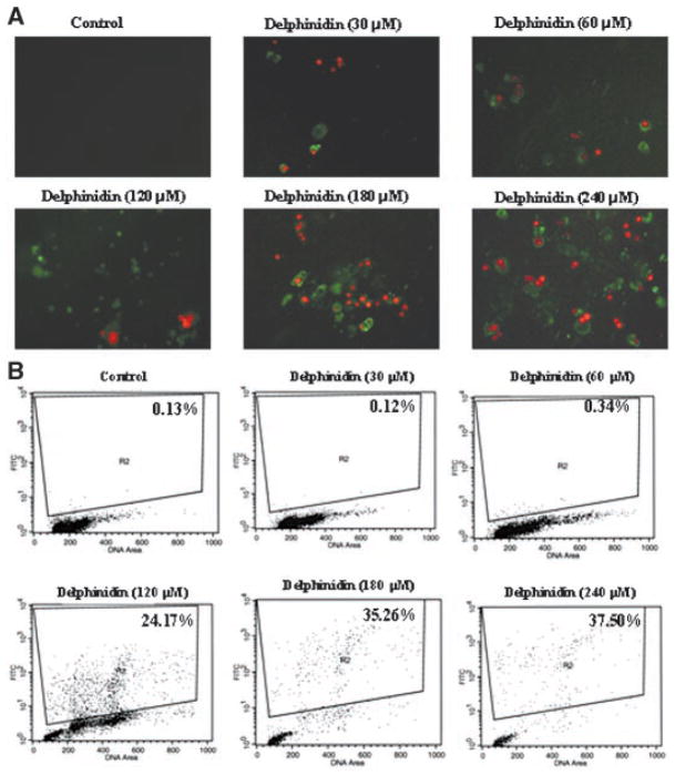
Figure 2.
Effect of delphinidin on apoptosis of HCT116 cells. (A) Fluorescence microscopy. The cells were treated with vehicle alone or specified concentration of delphinidin for 48 h. Annexin V-fluos staining kit was used for the detection of apoptosis and necrotic cells. Apoptosis and necrosis were detected by the kit according to the vendor’s protocol using a Zeiss Axiovert 100 microscope as described in Materials and Methods Section. The samples were excited at 330–380 nm, and the image was observed and photographed under a combination of a 400 nm dichroic mirror and the 420 nm high pass filter. Data from a typical experiment repeated three times with similar results are shown; Magnification 200×. (B). TUNEL Assay. The cells treated with delphinidin (30–240 μM; 48 h) by using apoptosis kit obtained from Phoenix Flow Systems (San Diego, CA) as per vendor’s protocol followed by flow cytometry. Cells showing deoxyuridine triphosphate fluorescence above that of control population, as indicated by the line in each histogram, are considered as apoptotic cells and their percentage population is shown in each box. The details are described in Materials and Methods Section.

