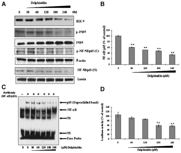
Figure 6.
Effect of delphinidin on IKKα, phosphorylation and degradation of IκBα and activation of NF-κB in HCT 116 cells. The cells treated with delphinidin (30–240 μM; 48 h) were harvested and nuclear lysate was prepared and protein was subjected to SDS-PAGE as detailed in Materials and Methods Section. (A) Immunoblot analysis of IKKα, IκBα, and NF-κB/p65. Equal loading of protein was confirmed by stripping the immunoblot and reprobing it for β-actin. The immunoblot shown here are representative of three independent experiments with similar results. (B) ELISA and (C) EMSA for NF-κB binding complex was demonstrated by anti-p65 antibody. (D) NF-κB transcriptional activity. At 48 h post-transfection with NF-κB luciferase plasmid, HCT116 cells were treated with the indicated concentration of delphinidin. Luciferase assay was performed as described in Materials and Methods Section.

