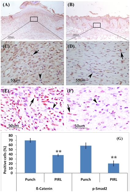
Figure 3. Less activation of β-Catenin and TGF- β signalling by using the PIRL laser.
Immunohistochemistry for β-Catenin and phospho-Smad2 staining (9 days post wounding) using Rabbit Anti-Beta-Catenin antibody shows an increased number of stained cells for β-Catenin and phospho-Smad2 in punch wounds. (A) and (C) for β-Catenin, (E) for p-Smad2 in punch wounds. (B) and (D) for β-Catenin, (F) for p-Smad2 in the PIRL laser wounds. While 70% (+/−5%) of cells in punch wounds (G) are positive for β-Catenin, only 38% (+/−3%) in the PIRL laser created wounds stained for β-Catenin. The ratios for pSmad2 positive cells are 58%(+/−8%) and 20%(+/−7%) for mechanical and PIRL laser results, respectively. Mean and 95% confidence interval are given. Arrows show positive cells for either β-Catenin or p-Smad2 staining while arrowheads show negative stained cells.

