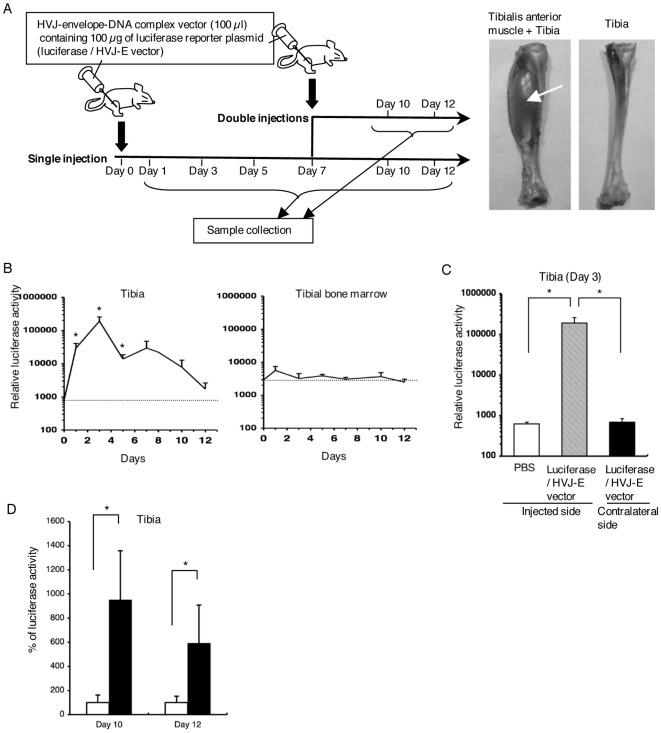
Figure 1. Gene expression in the interior of the bone by intramuscular administration of an HVJ-envelope-DNA complex.
(A) Experimental design of gene transfer. A luciferase/HVJ-E vector (100 µl) containing 100 µg luciferase plasmid DNA was carefully injected percutaneously into the proximal one-third of the tibialis anterior muscle of the right hindlimb. The first gene administration was on day 0, and the second on day 7. Samples were harvested on days 1, 3, 5, 7, 10, and 12 after the first administration of a luciferase/HVJ-E vector (each group: n = 7). Luciferase activity was measured using a luciferase assay system. Pictures show tibia with tibialis anterior muscle (arrow) and tibia from which the periosteum was thoroughly stripped to avoid any contamination with muscles. (B) Relative luciferase activity (RLU/mg protein) in the ipsilateral tibia (without periosteum), and bone marrow of the ipsilateral tibia. Error bars: SEM. Each group: n = 7. The dotted lines indicate the average levels of control samples, which were harvested at each time point after PBS injection (n = 6). Error bars: SEM. *p<0.05. (C) Relative luciferase activity (RLU/mg protein) in the tibia (without periosteum) on day 3 after gene transfer. Black bar: the activity in the contralateral tibia (n = 7), gray bar: the activity in the ipsilateral tibia (n = 7), white bar: the activity in the tibia after injection of PBS as a control (n = 6). Error bars: SEM. *p<0.05. (D) Comparison of luciferase activities of the ipsilateral tibia between single and repeated gene transfers on days 10 and 12. When luciferase activity in the ipsilateral tibia without a second gene transfer is considered as 100%, its activity in the ipsilateral tibia with second gene transfer on day 7 is shown as a percentage. Black bars: rats with the second gene transfer on day 7, white bars: rats without a second gene transfer. Error bars: SEM. Each group: n = 7. *p<0.05.

