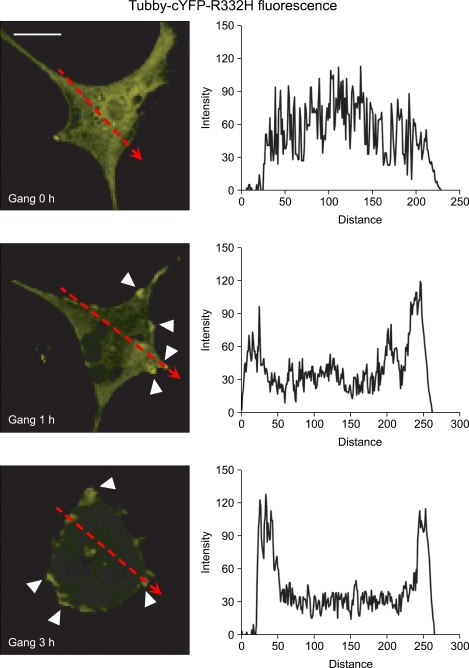
Figure 2.
Monitoring changes in PI(4,5)P2 levels induced by gangliosides in primary astrocytes. Primary astrocytes were transfected with a tubby-cYFP-R332H expression construct, a PI(4,5)P2-specific probe, using Amaxa Nucleofection. At 48 h posttransfection, cells were serum- starved and treated with 50 µg/ml gangliosides (Gang) for 0, 1, or 3 h. YFP fluorescence in the FITC channel was visualized using an LSM 710 confocal microscopy. Arrowheads indicate the localization of the tubby protein in the plasma membrane. Scale bar, 20 µm. YFP fluorescence intensities along the dotted line arrows in the cell images were analyzed using the Zeiss ZEN 2009 software. Their intensity profiles in the graphs show the translocation of tubby protein between the membrane and the cytosol.

