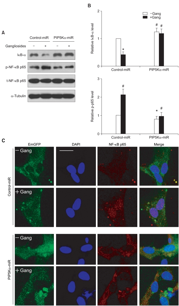
Figure 5.
Effects of PIP5Kα knockdown on ganglioside-stimulated NF-κB signaling. (A, C) Primary astrocytes were transfected with the miRNA expression plasmids and treated as described above. (A) After treatment with or without 50 µg/ml gangliosides for 15 min, cell lysates were prepared and changes in protein levels of IκB-α, phosphorylated and total NF-κB p65, and α-tubulin (a loading control) were examined by Western blot analysis using their specific antibodies. (B) IκB-α and phosphorylated NF-κB p65 levels in (A) were quantified relative to those in unstimulated negative control miRNA. Values are mean ± SEM. *P < 0.01 and #P < 0.05 compared with the unstimulated control miRNA. (C) Cells were stimulated with gangliosides under the same condition as (A) and then immunostained with a specific primary antibody against NF-κB p65, followed by an Alexa Fluor 594-conjugated secondary antibody. The transfected cells and the nuclei were visualized by EmGFP and DAPI staining, respectively. Cell images were obtained by an LSM 710 confocal microscopy under the laser filter sets (FITC, Rhodamine, and DAPI). Scale bar, 20 µm.

