Figure 1.
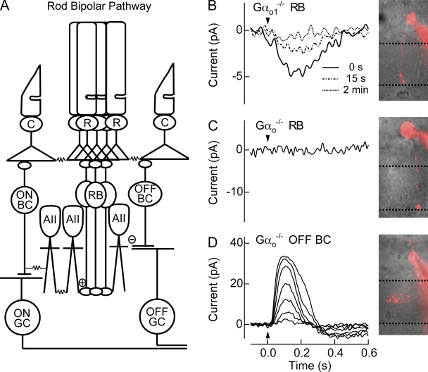
Rod bipolar responses are partially mediated by Gαo2. (A) Schematic of the mammalian rod bipolar pathway. Rod photoreceptors (R) synapse onto rod bipolar cells (RB), which in turn synapse onto AIIACs (AII). Signals from AIIACs, which are coupled to one another by Cx36 gap junctions (Deans et al., 2002), send light-driven signals to ON cone bipolar cells (ON BC) through gap junctions composed of Cx36 on the AII side, and make glycinergic (−) synapses with OFF cone bipolar cells (OFF BC). Each bipolar cells synapses with its respective ganglion cell (GC). Cone photoreceptors (C) are also depicted. (B) A representative Gαo1−/− rod bipolar cell visualized with Alexa 750 and the average flash response of 9 Gαo1−/− rod bipolar cells immediately after whole-cell break in (0 s), and 15 s and 2 min later. The flash strength was 15 Rh*/rod, a strength that saturates WT rod bipolar cells. (C) A representative Gαo−/− rod bipolar cell visualized with Alexa 750 did not generate light responses to flashes producing 32 Rh*/rod. In every rod bipolar cell tested from Gαo−/− mice, rod bipolar light responses were never observed. (D) To confirm viability within the retinal slice, a Gαo−/− Off-bipolar cell located near rod bipolar cell was visualized with Alexa 750, and displayed normal response families, indicating that the lack of rod bipolar responses was not due to the conditions of the retinal slice. Flash strengths were 0.5, 1.0, 2.0, 4.0, 8.0, 16, and 32 Rh*/rod.

