Figure 5.
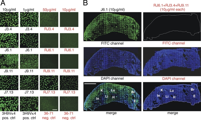
Immunofluorescence staining of HEp-2 cells and whole frozen sections of neonatal mouse with monoclonal antibodies and engineered revertants without somatic mutations. (A) Stains of fixed HEp-2 cells. R and red font color denote revertant. Note higher concentrations used for revertant antibodies. 3H9/Vκ4 and 36–71 served as positive and negative controls, respectively. The experiment was performed three times. Bar, 100 µm. (B) Stains of whole frozen sections of neonatal mice. Mutant antibody J6.1 served as positive control. Experimental section was stained with a mixture of revertant antibodies (RJ3.4, RJ9.11, and RJ6.1), each at a concentration equivalent to that of the positive control J6.1 mAb. Bound positive antibodies were detected with an FITC-coupled sheep anti–mouse IgG (γ-chain specific). Sections were counterstained with DAPI (blue) to highlight organs (In, intestine; Li, liver; K, kidney; Lu, lung; H, heart; Br, brain; E, eye). One of two experiments with similar results is shown. Bar, 3 mm.

