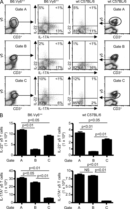
Figure 2.
Differential expression of IL-22 and IL-17A by Vγ6/Vδ1+ γδ T cells. (A) Representative density plots of intracellular IL-22 and IL-17A expression in γδ T cells isolated from the lung of WT C57BL/6 and B6.Vγ6+/+ mice treated with B. subtilis for 4 wk. γδ T cells expressing IL-17A or IL-22 were selected from a bivariate dot plot of CD3+/Cδ+ cells from the lymphocyte gate. The percentage of γδ T cells expressing either IL-17A or IL-22 is shown in each quadrant of the density plot. (B) Absolute number of IL-22– or IL-17A–expressing γδ T cells in the lung of B6.Vγ6+/+ and WT C57BL/6 treated with B. subtilis for 4 wk using different gating strategies labeled A, B, or C. Data represent at least five individual mice per experiment from at least three separate experiments (mean ± SD). NS, not significant. P > 0.05.

