
Figure 1.
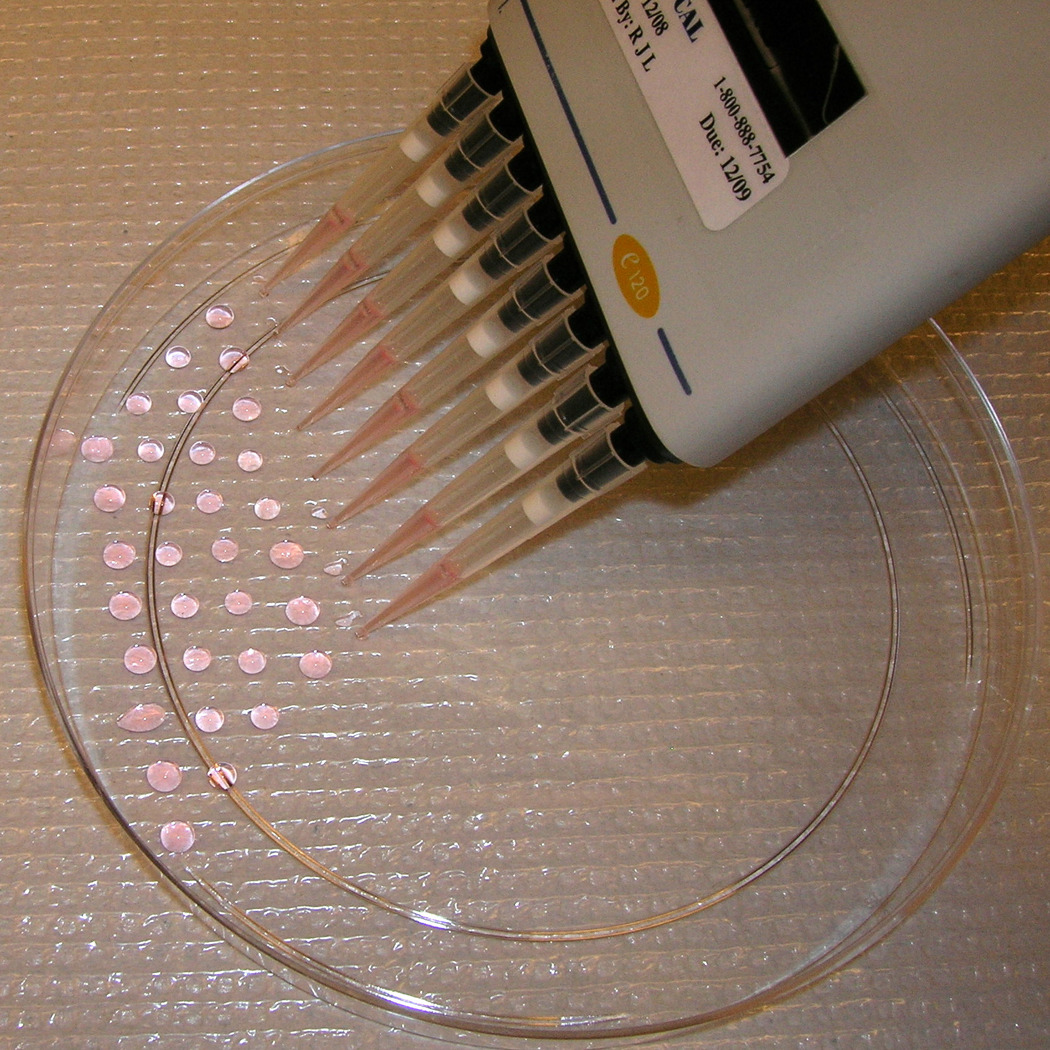
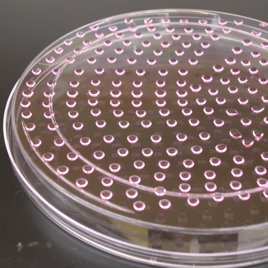
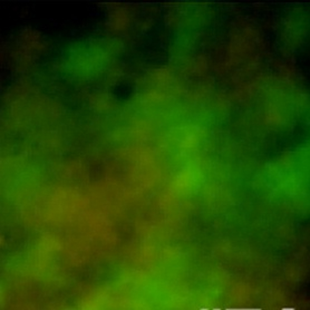
Embryoid body formation from hanging droplet. (a) Pipetting 11µL droplets of cells onto the bottom of a 15cm petri dish. (b) The dish is inverted to generate hanging droplets. The droplet density shown here (~250 droplets/plate) yields the maximum number of EBs per plate. (c) A representative area of beating eGFP+ cells within an EB.