
Figure 2.

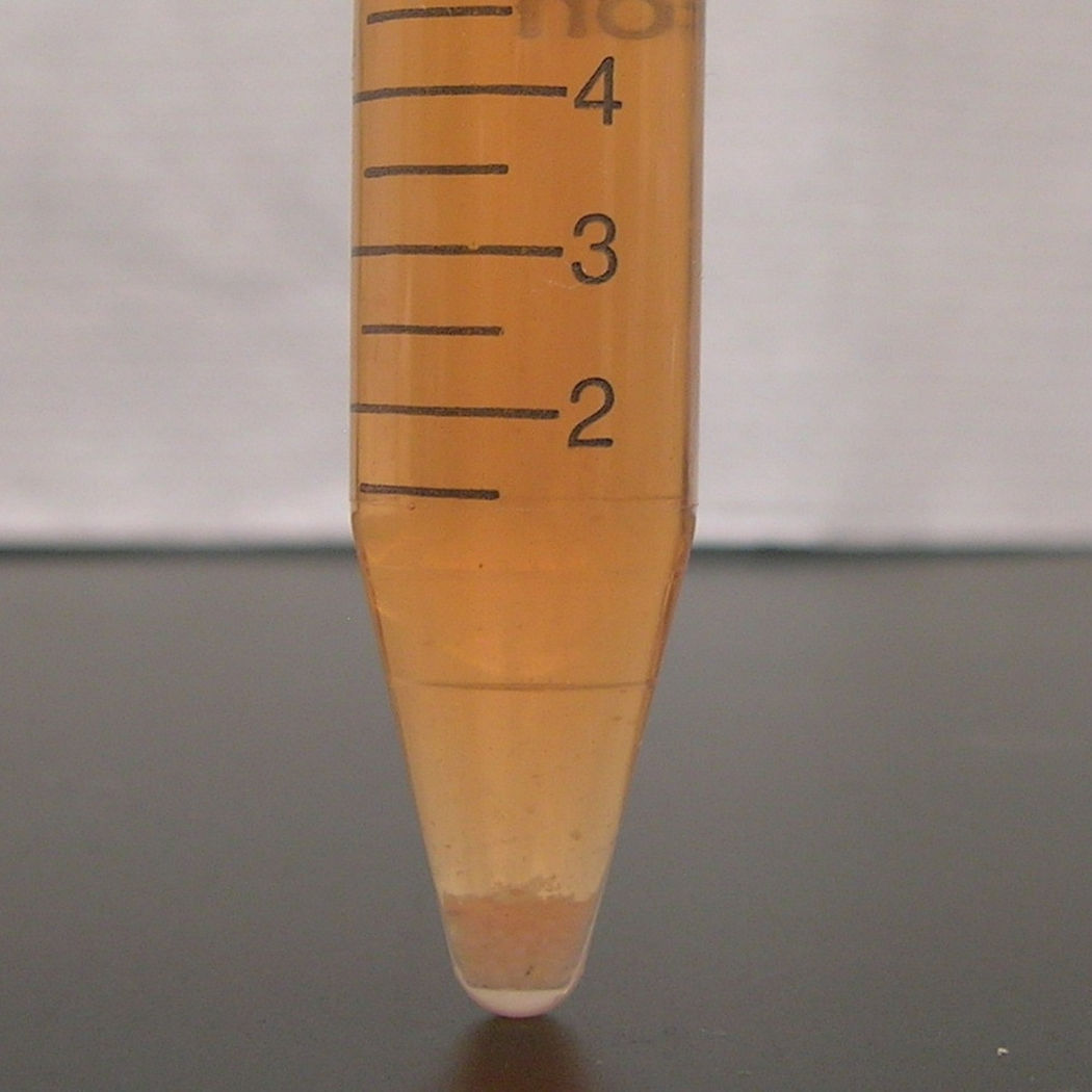
Harvesting EBs for isolation and analysis of cardiac progenitor cells. (a) After dissociation from the culture plate, EBs (indicated by arrows) appear as large particles in suspension. The plate shown is at Day 7 of differentiation. (b) The contents of the plate shown in (a) are transferred to a 15ml conical tube, and the EBs are allowed to settle to the bottom. Larger tubes (e.g. 50mL conical tube) may be used when pooling multiple plates.