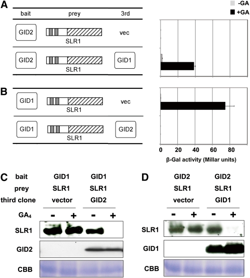
Figure 4.
GID1, GID2, and GA-Dependent Degradation of SLR1 in Yeast Cells.
(A) and (B) Interaction of SLR1-GID2 (A) and GID1-SLR1 (B) in yeast cells. Interaction of BD and AD fusion proteins in yeast cells with or without 10−4 M GA4 were scored using β-Gal activity (means ± sd; n = 3). Either GID2 or GID1 was used as bait, SLR1 was used as prey, and either GID1 or GID2 was expressed in yeast as a third clone.
(C) and (D) Accumulation of AD-HA-SLR1, HA-GID2, and HA-GID1 protein in yeast cells. Crude protein extracts from yeast grown in the absence or presence of 10−4 M GA4 were subject to immunoblot analysis and detected using the HA antibody for HA-SLR1 and HA-GID2, and anti-GID1 antibody for HA-GID1. The loading control of Coomassie blue (CBB) staining is shown in the bottom panels.
[See online article for color version of this figure.]

