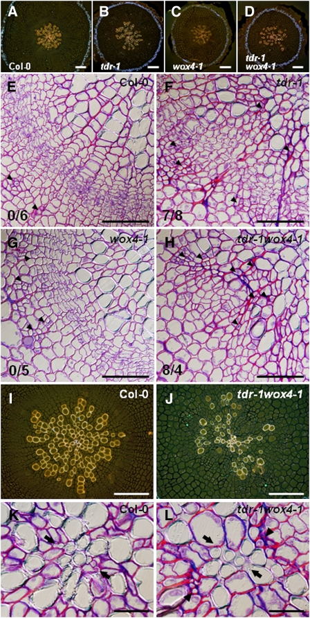
Figure 7.
Exhaustion of Vascular Stem Cells Caused Premature Termination of Secondary Growth.
(A) to (D) Overall radial structure of the 4-week-old hypocotyls.
(E) to (H) Magnifications of the cambial region located between the secondary phloem and xylem tissues. In contrast with the wild-type (E) and wox4-1 (G) plants, tdr (F) and tdr wox4 (H) plants had cavities in the outline of xylem tissues and the continuous cambium ring was disrupted. Some sieve elements (white arrowheads) were found at the bottom of the cavities ([F] and [H]). Numerals at left bottom show the number of cavities per the number of plants tested in (E) to (H).
(I) to (L) Central regions of the vascular cylinder in the wild type ([I] and [K]) and the most severely affected tdr-1 wox4-1 plant ([J] and [L]). In (K) and (L), the most central region of the vascular cylinder was magnified. Black arrows and arrowheads in (L) indicate the axis of primary xylem and primary phloem cells respectively. Stained by safranin-O and aniline blue, cells in xylem, phloem, and phellem (cork) show orange, green, and blue fluorescence, respectively, in (A) to (D), (I), and (J).
Bars = 100 μm in (A) to (D), (I), and (J), 50 μm in (E) to (H), and 20 μm in (K) and (L).

