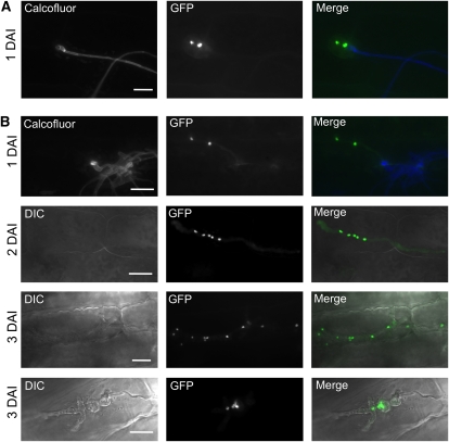
Figure 7.
Nuclear Distribution during Pathogenic Development of U. maydis Strains Expressing rbf1 and rbf1/clp1.
(A) Nuclear distribution in infection structures of a mixture of UKH184GN (a1 Δb::Plga2:rbf1 Pmig2_5:NLS-3eGFP) and UKH186GN (a2 Δb::Plga2:rbf1 Pmig2_5:NLS-3eGFP) expressing nuclear-localized GFP under the control of the plant-induced mig2-5 promoter. Calcofluor staining was used to visualize fungal growth on the leaf surface. Bulbous structures were formed directly after penetration of the host plant and invariably contained only the two nuclei of the dikaryotic hyphae formed after cell fusion. No progression of fungal development was observed at later time points of infection.
(B) Mixtures of UKH178GN (a1 Δb::Plga2:rbf1; Plga2:clp1 Pmig2_5:NLS-3eGFP) and UKH180GN (a2 Δb::Plga2:rbf1; Plga2:clp1 Pmig2_5:NLS-3eGFP), expressing nuclear-localized GFP during in planta growth. Hyphae infected the plant via appressorium-like structures, continued to proliferate inside the plant, and contained multiple nuclei. Hyphae were often found to contain uneven numbers and irregularly spaced nuclei. At 3 DAI, morphologically abnormal fungal hyphae (top panel, 3 DAI) or bulbous structures (bottom panel, 3 DAI) were frequently found that accumulated an increasing number of nuclei over time.
Bars = 20 μm.

